
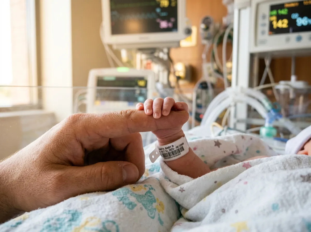
About 1 in every 3,500 babies is born with a condition called esophageal atresia, where the food pipe that connects the mouth to the stomach is either incomplete or missing a section entirely. In most cases, surgeons can connect the two ends. But in roughly 10% of those children, the gap is too wide. The upper and lower segments of the esophagus simply don't reach each other, and no amount of surgical stretching can bridge the distance. These children face years of feeding tubes, multiple surgeries, and borrowed tissue from other organs that was never designed to do the job of an esophagus.
A team of researchers at Great Ormond Street Hospital and University College London just demonstrated that there might be a better way. In a study published in Nature Biotechnology in March 2026, they took a donor pig's esophagus, stripped it of every cell until only the structural scaffold remained, repopulated that scaffold with the recipient's own muscle cells, and transplanted it into eight growing pigs. Every animal survived. Within six months, the engineered tissue had developed functional muscle, nerves, and blood vessels. The pigs swallowed normally, ate solid food, and grew at healthy rates. No immunosuppression was required.
It is the first time anyone has grown a functional esophagus in a lab and shown that it works in a living, growing animal. And it opens a door that regenerative medicine has been trying to push through for more than two decades.
The Problem With Borrowed Parts
To understand why this matters, you need to understand what long-gap esophageal atresia does to a child's life.
When surgeons can't connect the two ends of an interrupted esophagus, they have limited options. The most common replacement involves pulling up a section of the stomach and reshaping it into a tube, a procedure called a gastric conduit. It works, in the sense that food can pass through it, but stomach tissue isn't esophageal tissue. It doesn't contract in the same rhythmic waves that move food downward. It produces acid in a location where acid was never meant to be. Children who receive gastric conduits often struggle with reflux, aspiration, and difficulty swallowing for the rest of their lives.
Other approaches exist. The Foker procedure uses external traction sutures to gradually stretch the esophageal ends toward each other over weeks, but it requires multiple operations and extended sedation. A magnetic compression device approved by the FDA in 2017 uses opposing magnets to slowly draw the ends together, but it only works when the gap is relatively small. For children with truly long gaps, where the distance between segments can be several centimeters, none of these solutions provide what the child actually needs: a functioning esophagus made of esophageal tissue.
That is precisely what the Great Ormond Street team set out to build.
How You Grow an Esophagus
The technique rests on a principle that regenerative medicine researchers have been refining since 2008, when a team at the University of Minnesota demonstrated that you could strip a rat heart of all its cells, leaving only the extracellular matrix, the protein scaffolding that gives an organ its shape and mechanical properties, and then repopulate that ghost organ with new cells that would organize themselves into functional tissue.
The process is called decellularization, and it works because the extracellular matrix isn't just structural. It contains chemical signals, growth factors, and spatial cues that tell new cells where to go and what to become. A decellularized heart still looks like a heart. More importantly, it still tells cells how to be a heart.
The Great Ormond Street team applied this approach to a pig esophagus. They took a donor organ and carefully washed it with detergent solutions that dissolved the cellular material while preserving the underlying scaffold. What remained was a translucent tube with the exact architecture of an esophagus: the layers where muscle should be, the channels where nerves should run, the pathways where blood vessels should form.
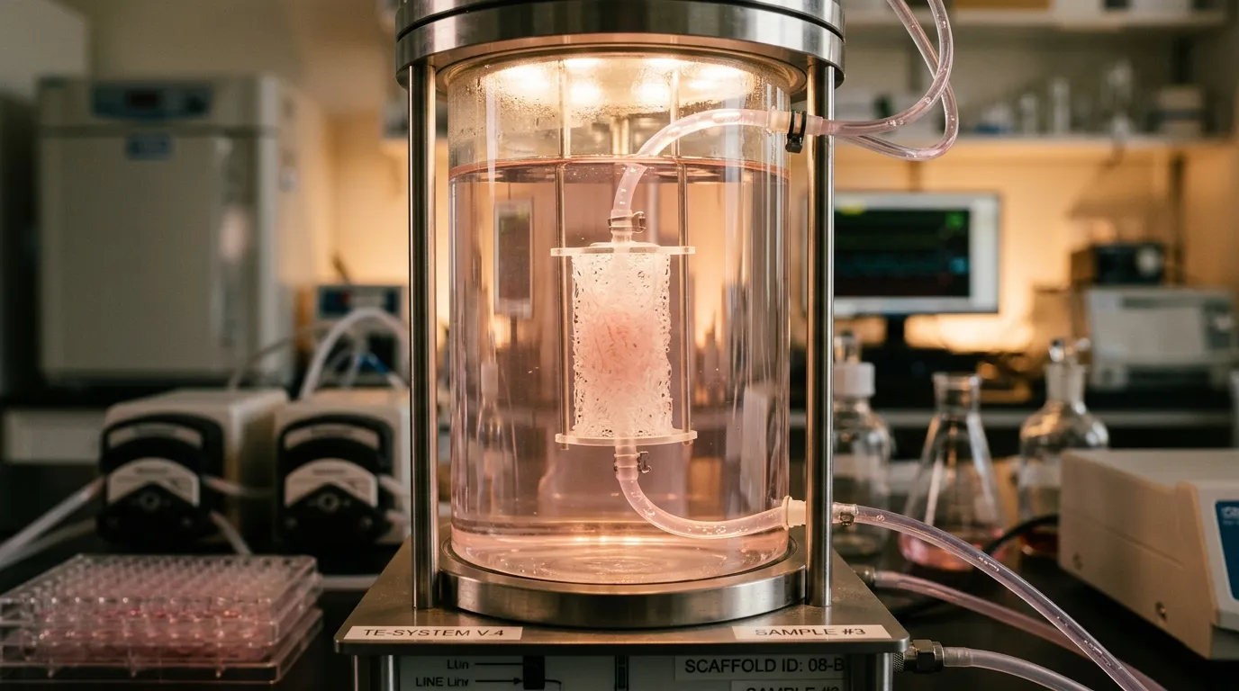
Next came recellularization. The researchers took a small muscle biopsy from each recipient pig, isolated the muscle cells, and expanded them in the laboratory over several weeks. They then injected those cells directly into the scaffold and placed the whole construct into a bioreactor, a chamber that pumps growth fluids through the tissue to simulate the biochemical environment of a living body. After one week in the bioreactor, the graft was ready for transplantation. The entire production process, from biopsy to implantable organ, took about two months.
What the Pigs Showed
All eight recipient pigs survived the initial 30-day critical window after transplantation. By three months, the grafts had fully integrated with the surrounding tissue. By six months, something remarkable had happened: the engineered esophagus had not only survived but matured. It had developed functional muscle capable of the peristaltic contractions that push food toward the stomach. New nerve fibers had grown into the tissue, providing the sensory and motor connections that coordinate swallowing. Blood vessels had formed throughout the graft, keeping it alive and nourished.
"Our grafts grew, matured and began to function like native tissue," said Dr. Natalie Durkin, the study's lead author. The pigs ate solid food, gained weight at normal rates, and showed no signs of immune rejection, despite receiving no immunosuppressive drugs at any point.
That last detail is especially significant. Organ transplants typically require a lifetime of immunosuppressive medication, which carries its own serious risks: increased vulnerability to infections, higher cancer rates, kidney damage over decades. Because the Great Ormond Street team used the recipient's own cells to repopulate the scaffold, the body recognized the engineered tissue as self rather than foreign. The immune system left it alone.
The results weren't perfect. Some of the pigs developed strictures, a narrowing of the engineered segment, that required endoscopic dilation to correct. But strictures are a manageable complication, far less burdensome than the chronic reflux and aspiration that plague children who receive gastric conduits.
Why the Esophagus Is Uniquely Difficult
Regenerative medicine has produced impressive results in other organs. Lab-grown bladders were implanted in human patients as early as 2006. Decellularized heart valves reached clinical use in 2001. Skin grafts grown from a patient's own cells have been commercially available for years. But the esophagus presents a set of challenges that those organs don't.
First, it moves. The esophagus is essentially a muscular tube that contracts in coordinated waves, and those contractions must be precisely timed and directional. A bladder mostly needs to hold fluid and contract. An esophagus needs to perform a complex mechanical sequence dozens of times a day. Building an organ that can do this requires not just muscle cells but the nerve connections that coordinate them.
Second, it grows. This technology is being developed for children, whose bodies are changing rapidly. An engineered organ that works in a newborn needs to still work in a ten-year-old. The Great Ormond Street team chose growing pigs as their animal model specifically to test whether the graft could keep pace with a developing body, and it did.
Third, it sits at a hostile intersection. The esophagus connects two very different environments: the throat, with its exposure to air, saliva, and bacteria, and the stomach, with its bath of hydrochloric acid. Any replacement tissue must withstand both, and it must do so while maintaining the tight cellular barriers that prevent acid from leaking into the chest cavity.
The fact that the engineered esophagus handled all three challenges in a growing animal model is what makes this study a genuine milestone rather than an incremental advance.
The Road to Human Trials
Professor Paolo De Coppi, who led the research, is cautiously optimistic. "We hope we can be successfully offering an engineered tissue alternative within five years," he said. But the path from pig model to pediatric operating room involves several steps that can't be rushed.
The team needs to refine the manufacturing process to make it reproducible and standardized. Every graft must perform identically, and the production timeline needs to be predictable enough that surgeons can plan operations around it. They also need to conduct further safety testing, including longer-term follow-up in animal models to confirm that the engineered tissue remains stable over years, not just months.
There are regulatory hurdles as well. Engineered organs occupy an unusual space in medical regulation. They aren't drugs, they aren't devices, and they aren't traditional transplants. Regulatory frameworks for these therapies are still being developed, and each country handles them differently.
Dr. Marco Pellegrini, who co-led the research, emphasized the clinical advantage that makes the regulatory effort worthwhile: the technology could enable construction of child-specific grafts "without the risk of rejection and without the need for long-term immunosuppression." For a child facing decades of life with a transplanted organ, eliminating immunosuppression isn't a convenience. It's a transformation.
The Bigger Picture
What makes this study resonate beyond pediatric surgery is what it proves about a broader idea: that you can take the architecture of one body, clean it of identity, rebuild it with the cells of another, and end up with something that functions as if it were always there. The scaffold remembers what it was. The new cells listen.
This principle, if it scales, has implications far beyond the esophagus. The same decellularization approach is being explored for kidneys, livers, lungs, and intestinal segments. Each organ brings its own complexity, its own cellular diversity, its own mechanical demands. But the underlying logic is the same: let biology's own blueprints guide the construction, rather than trying to engineer every detail from scratch.
For the roughly 18 children born each year in the UK with long-gap esophageal atresia, and the hundreds more worldwide, this research offers something that no existing treatment provides: the possibility of an esophagus that works like an esophagus, that grows with them, and that their bodies accept as their own. The gap in their anatomy may finally have a bridge that isn't borrowed from somewhere else.
Sources
- UCL News: Engineered Tissue Offers Hope for Babies Born with Missing Food Pipe Section
- Great Ormond Street Hospital: GOSH Charity-Funded Team Engineers First Lab-Grown Oesophagus
- News-Medical: Lab-Grown Food Pipe Offers Hope for Children with Esophageal Conditions
- Mirage News: Engineered Tissue Offers Hope for Missing Esophagus